Fig. 5: Cysts of Pneumocystis
jiroveci in Smear from Bronchoalveolar Lavage
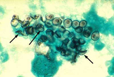
Cysts of Pneumocystis
jiroveci in lung tissue, Gomori methenamine silver stain method. The walls
of the cysts are stained black and often appear crescent shaped or like crushed
ping-pong balls. The intracystic bodies are not visible with this stain.
By Content Providers: CDC [Public domain].
Courtesy of the Centers for Disease Control and Prevention.

Microbiology Laboratory Manual by Gary E. Kaiser, PhD, Professor of Microbiology
is licensed under a Creative Commons Attribution 4.0 International License.
Last updated: September, 2017